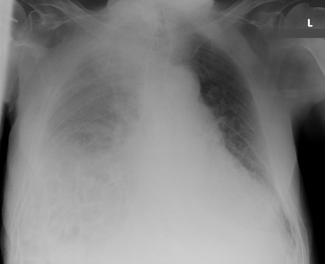
A patient was admitted to the department of traumatology for analgesic therapy of suspected fracture of lumbar vertebra after falling two days ago. CT was negative for fracture; just contusion was diagnosed.
The history of the patient includes chronic global heart failure, persistent atrial fibrillation on warfarin therapy, cholecystectomy, Chilaiditi syndrome. The patient complains of gradually worsening dyspnea at rest over the last six months. No other symptoms.
Chest X-ray performed because of SpO2 81 % while breathing ambient air and dyspnea at rest. The x-ray showed shadowing in the right hemithorax.
The patient was admitted to the intensive care unit

Chest X-ray performed during admission to a standard ward.
The next step after taking history and physical examination is performing POCUS:
